Butterfly Leg Under the Microscope
Butterfly Leg (Whole Mount), Lepidoptera
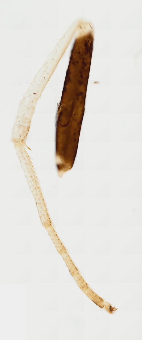
This is a butterfly leg mounted whole on a slide. It shows the jointed segments shared by all insect legs, plus scales and sensory hairs.
This whole mount slide preparation features the segmented thoracic leg of a butterfly (order Lepidoptera). The specimen clearly displays typical insect leg podomeres, including the elongated femur, tibia, and a five-segmented tarsus terminating in paired tarsal claws (ungues) and sensory structures. Fine, hair-like cuticular scales and sensory setae cover the amber-colored chitinous exoskeleton, serving tactile and chemoreceptive functions crucial for landing, perching, and substrate evaluation. Brightfield microscopy reveals the translucent cuticular boundaries, joint articulations, and localized pigmentation characteristic of adult lepidopteran leg morphology.
What to look for
- Femur: A long, thick segment near the body end of the leg.
- Tibia: The next long segment, joined to the femur at a bend.
- Tarsus: A chain of small segments that make up the foot.
- Tarsal claws: A pair of small, curved hooks at the tip of the leg for gripping.
- Scales and hairs: Tiny flat scales and fine hairs covering the amber-colored cuticle.
At each magnification
Low power (about 4x)
The full leg with its long segments and joints.
Medium power (about 10x)
The separate tarsal segments and the claws at the tip.
High power (about 40x)
Individual scales and sensory hairs on the leg surface.
Questions and answers
What are the parts of an insect leg?
An insect leg has the coxa and trochanter near the body, then a long femur, a long tibia, and a foot made of small segments called the tarsus, ending in claws. A butterfly leg shows these segments clearly.
Can butterflies taste with their feet?
Yes. Butterflies have taste receptors on their feet, which help them test whether a leaf is the right plant to lay eggs on.
- Category
- Invertebrates
- Scan magnification
- 20x
- Tags
- butterfly leg, Lepidoptera, whole mount, insect leg, tarsus, tarsal claw
Viewing this slide in class
This is a scan of a real prepared slide, not a simulation or a drawing. On StudyScope you can pan from the whole slide down to individual structures, always in focus, and measure them in real micrometers with the calibrated ruler. Present it live and every student screen follows your view and annotations. Students join with a room code on Chromebooks, iPads, or any browser, with no accounts.