House Fly Leg Under the Microscope
House Fly Leg (Whole Mount), Musca domestica
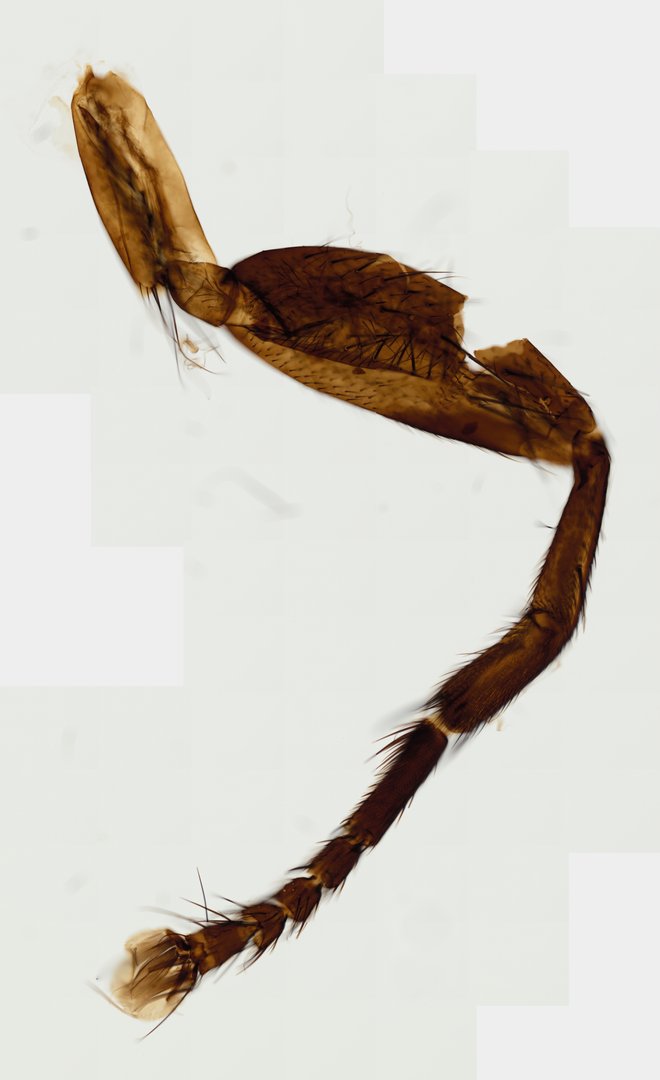
This is a house fly leg mounted whole and unstained. It shows all the standard insect leg segments, plus the sticky foot pads that let flies walk on walls and ceilings.
A whole mount (W.M.) preparation of a house fly leg. The slide displays typical dipteran leg segments including the coxa, trochanter, femur, tibia, and a five-segmented tarsus covered in sensory setae, terminating in a tarsal pretarsus equipped with claws and adhesive pulvilli.
What to look for
- Coxa and trochanter: Two short segments at the base where the leg joins the body.
- Femur: A long, thick segment after the trochanter.
- Tibia: A long segment after the femur.
- Tarsus: Five small segments forming the foot, covered with bristles.
- Claws and pulvilli: A pair of claws at the tip, with soft pads (pulvilli) beneath them.
At each magnification
Low power (about 4x)
The whole leg with its jointed segments and bristles.
Medium power (about 10x)
The five tarsal segments and the foot at the tip.
High power (about 40x)
The claws, the hairy pulvilli, and bristles along the segments.
Questions and answers
How do flies walk on the ceiling?
The tip of each fly leg has two claws and two soft pads called pulvilli. The pads are covered with tiny hairs that release a sticky fluid, which lets the fly grip smooth surfaces, while the claws grab rough ones.
What are the segments of a house fly leg?
A house fly leg has a coxa, trochanter, femur, tibia, and a tarsus of five segments. The last segment carries the claws and pads.
- Category
- Invertebrates
- Scan magnification
- 20x
- Stain
- Unstained
- Tags
- House fly leg whole mount, House Fly Leg W.M., Musca domestica, House fly, Diptera, Muscidae
Viewing this slide in class
This is a scan of a real prepared slide, not a simulation or a drawing. On StudyScope you can pan from the whole slide down to individual structures, always in focus, and measure them in real micrometers with the calibrated ruler. Present it live and every student screen follows your view and annotations. Students join with a room code on Chromebooks, iPads, or any browser, with no accounts.