Locust Testis Cross Section Under the Microscope
Locust Testis (Transverse Section), Locusta migratoria
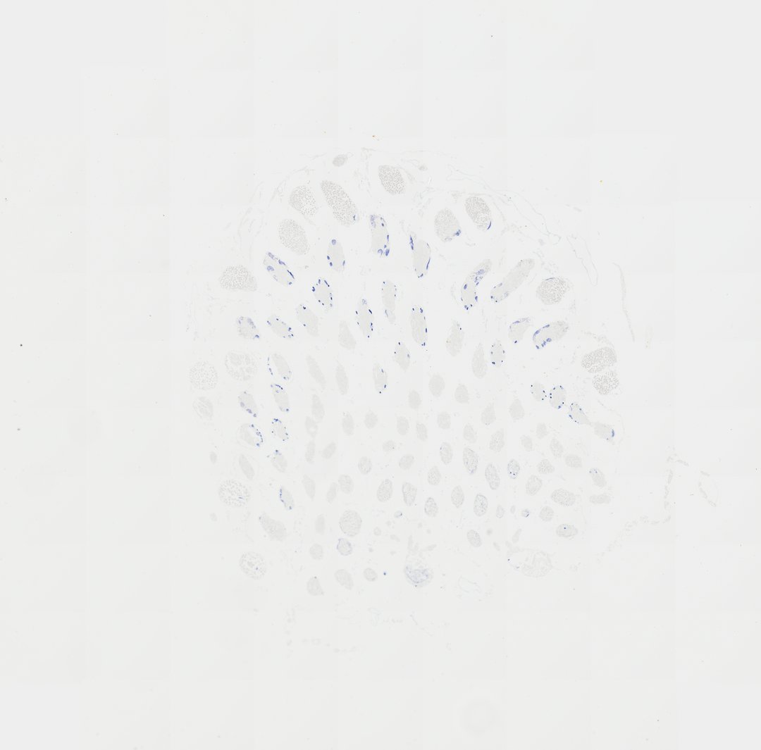
This is a cross section of a locust testis stained with hematoxylin. Grasshopper and locust testes are classic slides for meiosis because they contain many cells at different stages of making sperm.
A transverse section (T.S.) of a locust testis (labeled Locusts testis T.S.). The slide displays cross-sections through multiple testicular follicles (testioles), revealing various stages of spermatogenesis within the germinal cysts.
What to look for
- Testis follicles: Round or oval tubes, cut across, that make up the testis.
- Cysts: Groups of germ cells inside the follicles, all at about the same stage.
- Dividing cells: Cells with dark, condensed chromosomes, some caught in meiosis.
- Spermatids: Small cells with compact nuclei, formed after meiosis.
- Sperm bundles: Groups of long, thin, dark-stained sperm.
At each magnification
Low power (about 4x)
Several round follicles packed together.
Medium power (about 10x)
Cysts of cells at different stages within the follicles.
High power (about 40x)
Chromosomes in dividing cells, stained dark blue to purple by hematoxylin.
Questions and answers
Why is locust or grasshopper testis used to show meiosis?
The testes are full of cells making sperm, and each cyst holds many cells at the same stage. Their chromosomes are large and easy to stain, so different stages of meiosis can be seen on one slide.
What is the difference between mitosis and meiosis on a slide?
Mitosis makes two identical cells and is seen in growing tissue like onion root tips. Meiosis makes sex cells with half the chromosomes and is seen in testes and anthers, where you may spot paired chromosomes and groups of four cells.
- Category
- Invertebrates
- Scan magnification
- 20x
- Stain
- Hematoxylin
- Tags
- Locusts testis T.S., Locust testis t.s., Locust testis transverse section, Locust testis cross section, Locust testis, Locusta migratoria
Viewing this slide in class
This is a scan of a real prepared slide, not a simulation or a drawing. On StudyScope you can pan from the whole slide down to individual structures, always in focus, and measure them in real micrometers with the calibrated ruler. Present it live and every student screen follows your view and annotations. Students join with a room code on Chromebooks, iPads, or any browser, with no accounts.