Dragonfly Wing Under the Microscope
Dragonfly Wing (Whole Mount), Anisoptera
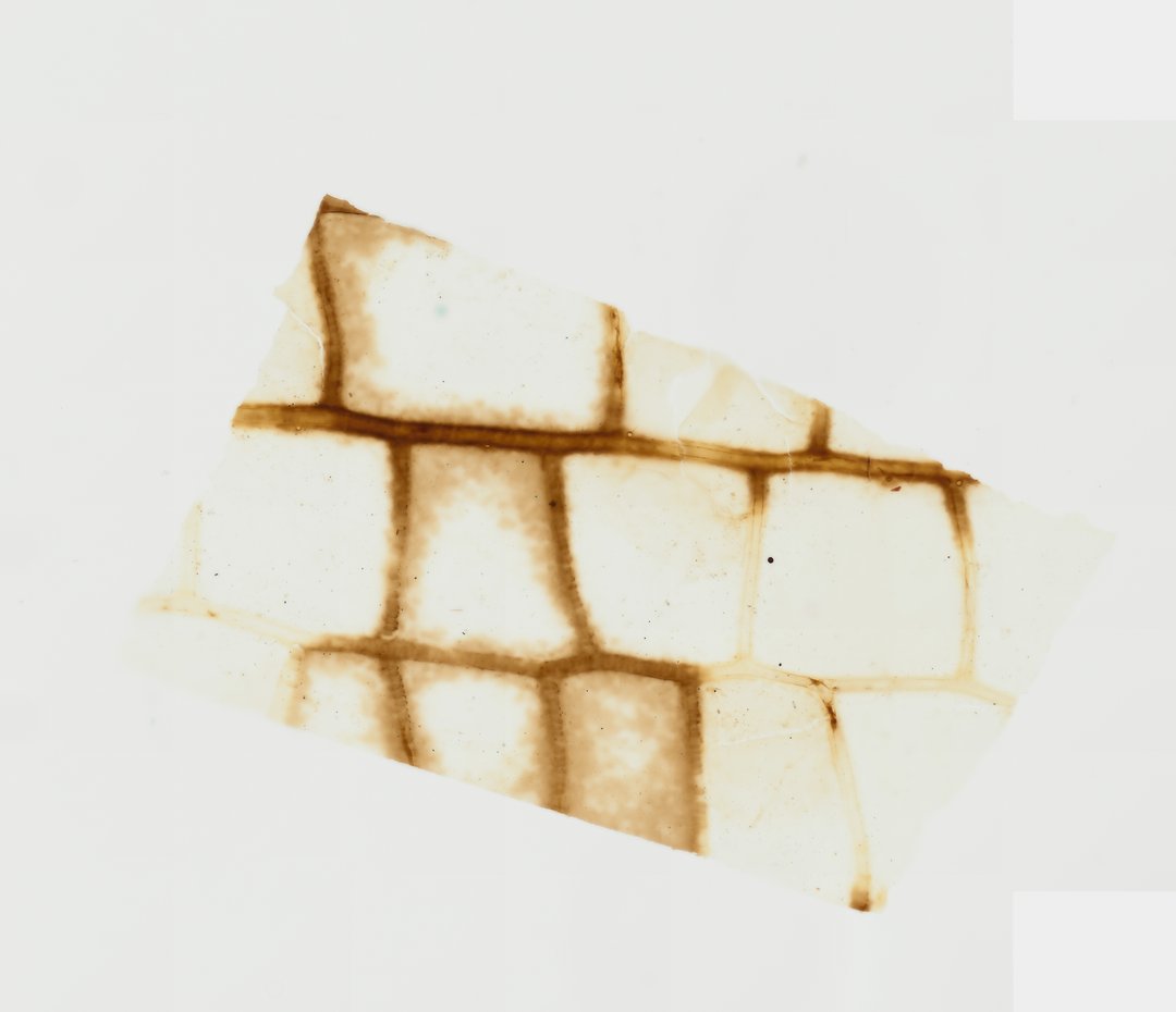
This slide shows part of a dragonfly wing mounted whole. The network of veins makes a light but strong frame, and it is a nice example of how structure supports function.
This whole mount preparation presents a section of a dragonfly wing (order Odonata, suborder Anisoptera), detailing its specialized biomechanical architecture. The clear, membranous cuticular lamina is reinforced by a complex framework of sclerotized veins forming a lattice of closed cells. Visible within the section are longitudinal major veins interconnected by transverse cross-veins, creating a lightweight yet rigid structural grid optimized for high-performance flight aerodynamics and torsional flexibility. Under brightfield illumination, the dark amber pigmentation of the chitinous veins contrasts sharply against the hyaline wing membrane, allowing precise observation of wing venation patterns critical for odonate identification and insect structural biology.
What to look for
- Wing membrane: The thin, clear sheet between the veins.
- Longitudinal veins: Main veins running along the length of the wing, dark amber in color.
- Cross veins: Many short veins connecting the main veins.
- Cells: The small enclosed areas of membrane between veins, forming a net-like pattern.
At each magnification
Low power (about 4x)
A net of dark veins across a clear wing.
Medium power (about 10x)
The pattern of main veins and cross veins forming many small cells.
High power (about 40x)
The surface of individual veins and the texture of the membrane.
Questions and answers
What does a dragonfly wing look like under a microscope?
A dragonfly wing is a thin, clear membrane held in a dense network of dark veins. The main veins run along the wing and many short cross veins link them, forming lots of small cells.
Why do dragonfly wings have so many veins?
The network of veins makes the wing stiff and strong while keeping it light. This helps dragonflies hover, turn quickly, and even fly backward.
- Category
- Invertebrates
- Scan magnification
- 20x
- Tags
- dragonfly wing, Anisoptera, Odonata, whole mount, wing venation, chitinous veins
Viewing this slide in class
This is a scan of a real prepared slide, not a simulation or a drawing. On StudyScope you can pan from the whole slide down to individual structures, always in focus, and measure them in real micrometers with the calibrated ruler. Present it live and every student screen follows your view and annotations. Students join with a room code on Chromebooks, iPads, or any browser, with no accounts.