Earthworm Cross Section Under the Microscope
Earthworm (Cross Section), Lumbricus terrestris
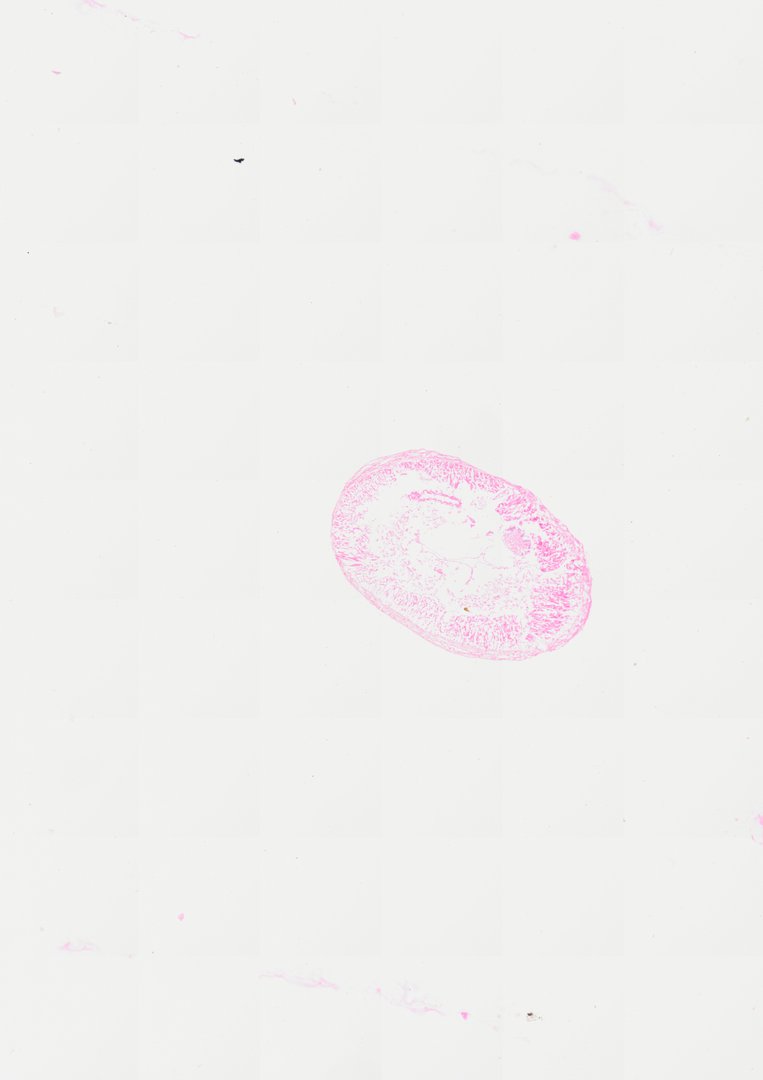
This is a cross section of an earthworm stained with H&E. It shows the classic body plan of a segmented worm, with a tube (the gut) inside a tube (the body wall) and a fluid-filled cavity between them.
A cross section (C.S.) of an earthworm showcasing typical coelamate annelid structural organization. The outer wall features a thin cuticle lining an epidermal layer, followed by well-developed circular muscle and thick longitudinal muscle bands organized in feather-like bundles. The central coelomic cavity surrounds an intestinal tract with a distinct dorsal typhlosole fold and outer chloragogen cell layer, alongside major longitudinal blood vessels and ventral nerve cord components.
What to look for
- Cuticle and epidermis: A thin outer covering over a single layer of skin cells.
- Circular muscle: A thin layer of muscle just inside the epidermis.
- Longitudinal muscle: A thicker layer of muscle, pink with H&E, arranged in feather-like bundles.
- Coelom: The open body cavity between the body wall and the intestine.
- Intestine and typhlosole: The central gut, with a fold called the typhlosole dipping in from the top.
- Ventral nerve cord: A small round cord along the bottom of the body cavity.
At each magnification
Low power (about 4x)
A round section with a muscular outer wall and a central gut with an inward fold.
Medium power (about 10x)
The two muscle layers, the coelom, and the blood vessels above and below the gut.
High power (about 40x)
Cells lining the intestine, the chloragogen cells around it, and nuclei stained purple.
Questions and answers
What are the parts of an earthworm cross section?
An earthworm cross section shows the cuticle and epidermis, circular and longitudinal muscle layers, the coelom (body cavity), and the intestine with its typhlosole fold. You can also find the dorsal blood vessel above the gut and the ventral blood vessel and nerve cord below it.
What is the typhlosole?
The typhlosole is a fold of the intestine wall that hangs down into the gut. It increases the surface area for absorbing food, similar to how villi work in the human small intestine.
How do earthworms move using two muscle layers?
When circular muscles contract, the worm gets long and thin. When longitudinal muscles contract, it gets short and fat. Alternating these, along with small bristles that grip the soil, lets it crawl.
- Category
- Invertebrates
- Scan magnification
- 20x
- Stain
- H&E
- Tags
- Earthworm cross section, Earthworm C.S., Lumbricus terrestris, Earthworm, Annelida, Oligochaeta
Viewing this slide in class
This is a scan of a real prepared slide, not a simulation or a drawing. On StudyScope you can pan from the whole slide down to individual structures, always in focus, and measure them in real micrometers with the calibrated ruler. Present it live and every student screen follows your view and annotations. Students join with a room code on Chromebooks, iPads, or any browser, with no accounts.