Hirudo Cross Section Under the Microscope
Hirudo (Transverse Section), Hirudo medicinalis
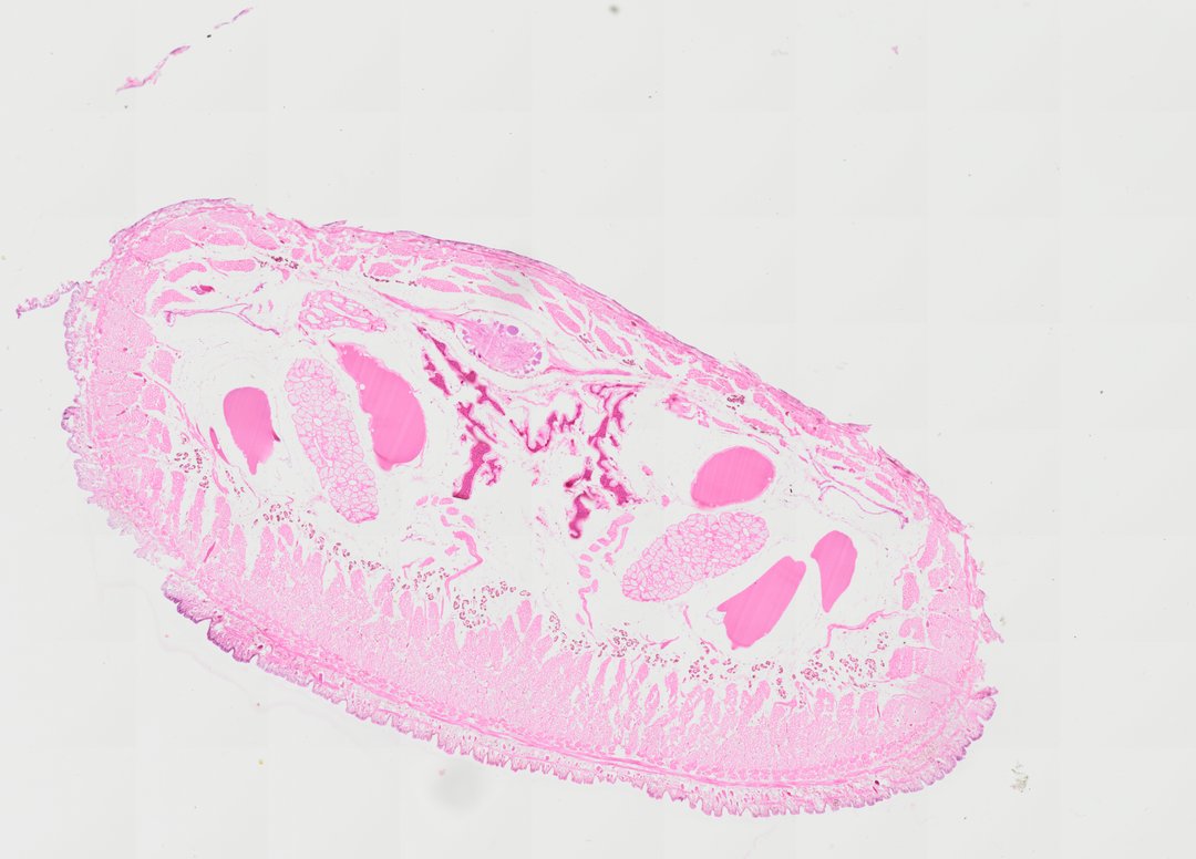
This is a cross section of the medicinal leech, Hirudo, stained with H&E. Leeches are segmented worms like earthworms, but their body is packed with muscle and tissue instead of an open body cavity.
A transverse section (T.S.) of the medicinal leech, Hirudo medicinalis, demonstrating characteristic annelid morphological organization. The section reveals an outer cuticle overlying a single-layered epidermis with abundant mucosal gland cells. Beneath the body wall, prominent circular and powerful dorsoventral and longitudinal muscle bundles are embedded within a dense, reduced coelomic parenchymal matrix (botryoidal tissue). Large, distinct coelomic sinuses, lateral blood vessels, and crop diverticula pockets filled with homogeneous fluid are visibly distributed across the internal parenchymal space.
What to look for
- Cuticle and epidermis: A thin outer covering over a single layer of skin cells, with gland cells.
- Muscle layers: Bands of circular, diagonal, and longitudinal muscle, stained pink, forming a thick body wall.
- Dorsoventral muscles: Muscle strands crossing from the top to the bottom of the body, which help flatten it.
- Botryoidal tissue: Packed tissue filling much of the space inside the body wall.
- Crop: Large pouches of the gut used to store blood meals.
At each magnification
Low power (about 4x)
A flattened oval section with a thick muscular wall and large gut pouches.
Medium power (about 10x)
The muscle layers and the tissue filling the body.
High power (about 40x)
Individual muscle fibers and gland cells in the epidermis, with purple nuclei.
Questions and answers
How is a leech different from an earthworm in cross section?
An earthworm has a large open body cavity (coelom) around its gut. In a leech, the coelom is mostly filled in with muscle and tissue, and leeches have no bristles (setae) for gripping the soil.
Why are leeches used in medicine?
Medicinal leeches release chemicals in their saliva that stop blood from clotting. Doctors sometimes use them to help blood flow in reattached fingers or skin grafts.
- Category
- Invertebrates
- Scan magnification
- 20x
- Stain
- H&E
- Tags
- Hirudo transverse section, Hirudo T.S., Hirudo medicinalis, Leech, Annelida, Hirudinea
Viewing this slide in class
This is a scan of a real prepared slide, not a simulation or a drawing. On StudyScope you can pan from the whole slide down to individual structures, always in focus, and measure them in real micrometers with the calibrated ruler. Present it live and every student screen follows your view and annotations. Students join with a room code on Chromebooks, iPads, or any browser, with no accounts.