Oyster Gill Cross Section Under the Microscope
Oyster Gill (Cross Section), Crassostrea virginica
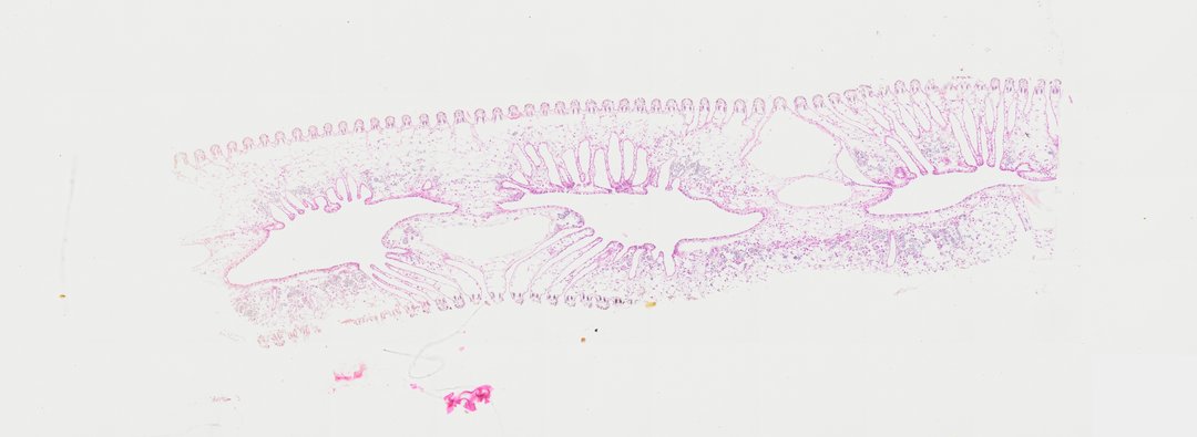
This is a cross section of an oyster gill stained with H&E. Oyster gills are used for both breathing and filter feeding, trapping tiny food particles from the water.
A cross section (c.s.) of an oyster gill (labeled Oyster gill c.s.). The slide demonstrates the complex bivalve demibranch architecture, showing w-shaped ciliated gill filaments, ostia, and internal water tubes involved in both respiration and filter feeding.
What to look for
- Gill filaments: Many thin, closely spaced filaments arranged in rows, forming a folded pattern.
- Cilia: Tiny hair-like structures along the surface of the filaments that move water and food.
- Water tubes: Spaces inside the gill where water flows after passing between the filaments.
- Connecting tissue: Pink bridges of tissue that join neighboring filaments.
At each magnification
Low power (about 4x)
A folded, W-shaped pattern of gill tissue.
Medium power (about 10x)
Rows of filaments and the water tubes between them.
High power (about 40x)
Cells lining the filaments, with purple nuclei and a fringe of cilia.
Questions and answers
How do oysters use their gills to eat?
Oysters are filter feeders. Cilia on their gills pull water through, trap tiny food particles in mucus, and move the food toward the mouth.
What does an oyster gill look like under a microscope?
An oyster gill looks like rows of thin filaments folded into a W shape, with water tubes inside. The filaments are lined with cilia that keep water moving.
- Category
- Invertebrates
- Scan magnification
- 20x
- Stain
- H&E
- Tags
- Oyster gill c.s., Oyster gill cross section, Oyster gill t.s., Oyster gill transverse section, Oyster gill, Crassostrea virginica
Viewing this slide in class
This is a scan of a real prepared slide, not a simulation or a drawing. On StudyScope you can pan from the whole slide down to individual structures, always in focus, and measure them in real micrometers with the calibrated ruler. Present it live and every student screen follows your view and annotations. Students join with a room code on Chromebooks, iPads, or any browser, with no accounts.