Planaria Cross Section Under the Microscope
Planaria (Transverse Section), Dugesia
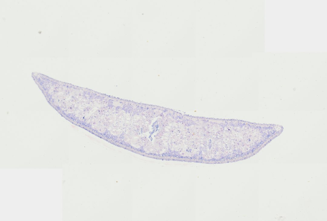
This is a cross section of a planarian, a small freshwater flatworm. Planarians have no body cavity and a branched gut, and they are famous for their ability to regrow lost body parts.
A transverse section (T.S.) of a planarian flatworm demonstrating dorosventrally flattened triploblastic anatomy. The section reveals an outer ciliated epidermis lined with rhabdites, an underlying loose parenchymal tissue network, and muscle layers arranged in circular and longitudinal orientations. Centrally, branches of the gastrovascular cavity with digestive gastrodermal cells and luminal spaces are clearly identifiable, characteristic of acoelomate Platyhelminthes morphology.
What to look for
- Flattened body: A section that is wide and thin, since flatworms are flattened from top to bottom.
- Epidermis: A single outer layer of cells, with cilia on the underside for gliding.
- Muscle layers: Thin layers of muscle just under the epidermis.
- Parenchyma: Loose tissue filling the body, since planarians have no body cavity.
- Gut branches: Several spaces lined with cells, which are cross sections of the branched gut.
At each magnification
Low power (about 4x)
A wide, flat oval section with several small cavities inside.
Medium power (about 10x)
The gut branches and the tissue filling the space around them.
High power (about 40x)
Cells of the epidermis and the cells lining the gut.
Questions and answers
What does a planarian cross section look like under a microscope?
A planarian cross section is flat and oval, with an outer epidermis, thin muscle layers, and loose tissue filling the body. Several small cavities are cut through the branches of its gut.
What does acoelomate mean?
Acoelomate means having no body cavity. In flatworms like planaria, the space between the gut and the body wall is filled with solid tissue instead of a fluid-filled cavity.
Why are planarians used in science?
Planarians can regrow a whole body from a small piece. Scientists study them to learn how stem cells rebuild tissues.
- Category
- Invertebrates
- Scan magnification
- 20x
- Tags
- Planaria transverse section, Planaria T.S., Planaria, Dugesia, Platyhelminthes, Turbellaria
Viewing this slide in class
This is a scan of a real prepared slide, not a simulation or a drawing. On StudyScope you can pan from the whole slide down to individual structures, always in focus, and measure them in real micrometers with the calibrated ruler. Present it live and every student screen follows your view and annotations. Students join with a room code on Chromebooks, iPads, or any browser, with no accounts.